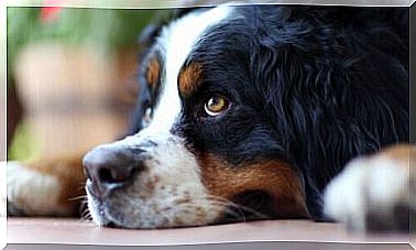
Scintigraphy In Veterinary Medicine: What Is It And How Does It Work?
The scintigraphy is a diagnostic technique of nuclear medicine. It uses radioactive isotopes to recognize and follow various types of pathologies.
This term also indicates the specialty that directly deals with the study and application of this methodology. Let’s try to better understand what veterinary scintigraphy is .
How does scintigraphy work?
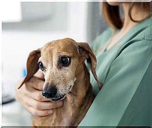
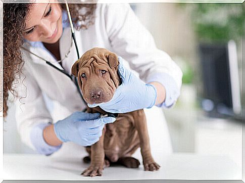
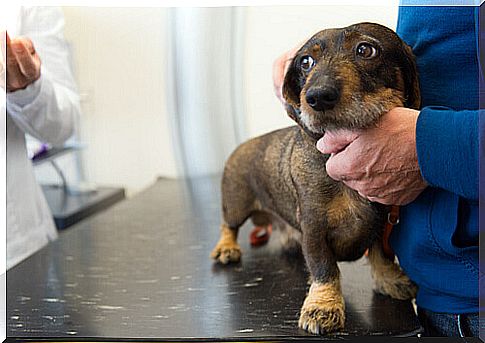
The patient who has to perform a scan, whether human, dog or cat, receives a controlled dose of reactive isotopes ( radioisotopes ).
These substances can be administered orally or injected directly into the blood intravenously.
The type of isotope used depends on the purpose of the study and the type of organ to be analyzed. Iodine-131 is used with labeled albumin to analyze the brain. When combined with 198 colloidal gold, it is used to examine the liver. Chromium 51 is applied to study the spleen.
These substances move through the bloodstream to reach certain organs. Then, a machine called the scintillation counter (or scintillator) covers the entire contour of the patient’s body with its camera.
This device detects the gamma rays resulting from the reaction of the reactive isotopes supplied to the patient. The emission of these rays derives from the release of radioactive particles derived from the spontaneous decay of radioisotopes.
The computer connected to the scintillator records and transmits the behavior of the isotopes in the form of images and action curves. This allows you to obtain a high-quality overview of the internal structures of the organism, such as tissues and walls of different organs.
Applications of scintigraphy in veterinary medicine
Regarding the applications, the scintigraphy technique offers morphological information (on the anatomy and structure of the organs) and functional information.
It allows to recognize different pathologies in an early way. Promotes early diagnosis and a better prognosis for the patient.
In fact, in degenerative diseases its action is crucial for survival.
Thanks to its high sensitivity, scintigraphy is very effective in diagnosing benign and malignant tumors. Normally, it is complementary to other imaging techniques , such as tomography and ultrasound scans.
In addition to oncology, this technique is also used in the following medical areas :
- Gastroenterology
- Endocrinology.
- Cardiorespiratory.
- Neurology.
- Nephrology.
- Urology.
Veterinary scintigraphy and oncology
Compared to bone, scintigraphy is used to analyze and evaluate first and second degree tumors. It is effective in detecting bone-type lesions and detecting metastases caused by breast cancers.
As for its application in the bone marrow, it is effective for recognizing aplasia, hypoplasia, hyperplasia and abnormal masses.
It has also been shown to be useful in effectively diagnosing the existence of malignant tumors and lesions in lymphatic and breast tissue, and in detecting the existence of topical adenopathy.
On the other hand, the other modality, called lymphocentography, is mainly used to locate the sentinel lymph node in cases of melanoma.
When a Gallium 67 isotope is administered it will be possible to identify non-visible abscesses and immunological disorders in patients with lymphoma.
Veterinary endocrinology scintigraphy
In veterinary endocrinology, the main use of scintigraphy is associated with the diagnosis of thyroid cancer.
This technique allows to evaluate the morphology of the gland and to detect disturbances in its structure and functioning.
It is also effective in recognizing the presence of malignant nodules, hyperplasia, congenital hypothyroidism, and nodular disease.
Therefore, the patient should discontinue the daily administration of levothyroxine or methylmercaptoimidazole so as not to interfere with the quality of the study.
Finally, endocrine scintigraphy also investigates the pancreatic area and is able to indicate hyperplasia, neoplasms and insulinomas.
Veterinary gastroenterology scintigraphy
It is used to carry out analyzes in salivary, hepatobiliary and transcolonic cells. Its high sensitivity allows you to diagnose the following conditions:
- Analysis of inflammation and sialolithiasis (salivary region).
- Verification of the function of the gallbladder, hepatocytes and excretory passages of the liver, mainly in patients diagnosed with gallbladder and liver disease.
- Perception and measurement of portosystemic shunt fractionation in the colon region.
Cardiorespiratory scan
- Lung diagnostics : Detection and monitoring of pulmonary embolism (PE), as well as verification of pulmonary pressure in the presence of cases of derivation of congenital heart disease.
- Myocardial analysis : identifies the perfusion of different areas of the myocardium, heart disease and arrhythmias and also differentiates clots from abnormal masses.
- Angiography : Checks the flow of blood circulation and identifies the location of thromboemboli and kidney function.
Scintigraphy also makes it possible to investigate and recognize tumors and infections in the brain, which is why its use as a diagnostic tool in veterinary neurology is high.